Article No. VK-MED08
MED08 Ultrasound fetus phantom
Simulation of biometric measurements as carried out during pregnancy, Examination of image artefacts
- Subject matter of the experiment
- Theoretical and practical aspects of the experiment
- Results
- Equipment
- Related Experiments
With the aid of the ultrasound fetal phantom, biometric measurements can be simulated, as they are carried out during a pregnancy examination. Furthermore, image artifacts are examined in the ultrasound image, which are generated by phenomena of ultrasonic propagation or by steps of signal and image processing.
Keywords: Ultrasound diagnostics, sonography, B-scan, fetometry, transmitting power, total gain, TGC, dynamic range, measurement depth, image artifacts
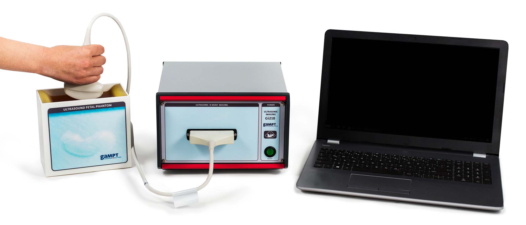
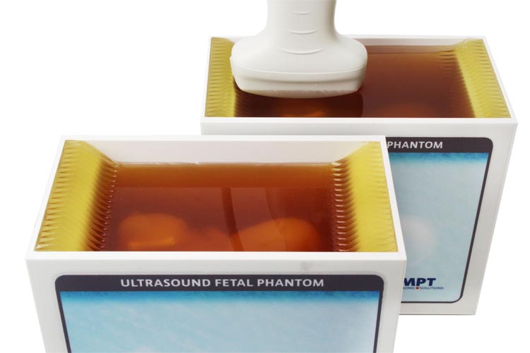
Sonographc examinations during pregnancy are standard today. In addition to assessing the position of the child and the placenta, the amount of amniotic fluid or the heartbeat, the fetus is also measured (fetometry = biometry of the fetus). Depending on the age of the fetus different biometric variables (head circumference, femur length, etc.) are determined. With the help of these quantities the age of a fetus can be estimated. If the age is known, the physician can use the measurements to assess whether the fetus has developed according to his age or whether there may be malformations or developmental delays.In combination with the GS200i or Gi210 echoscope and a convex multi-element ultrasound probe for the abdominal region, the fundamentals of sonographic fetometry can already be mediated in pre-clinical training at GAMPT‘s ultrasonic fetus phantom.Note: Please use only water as coupling medium (no gel). For this purpose, the water should be poured into the upper part of the phantom so that the transducer can be immersed well.
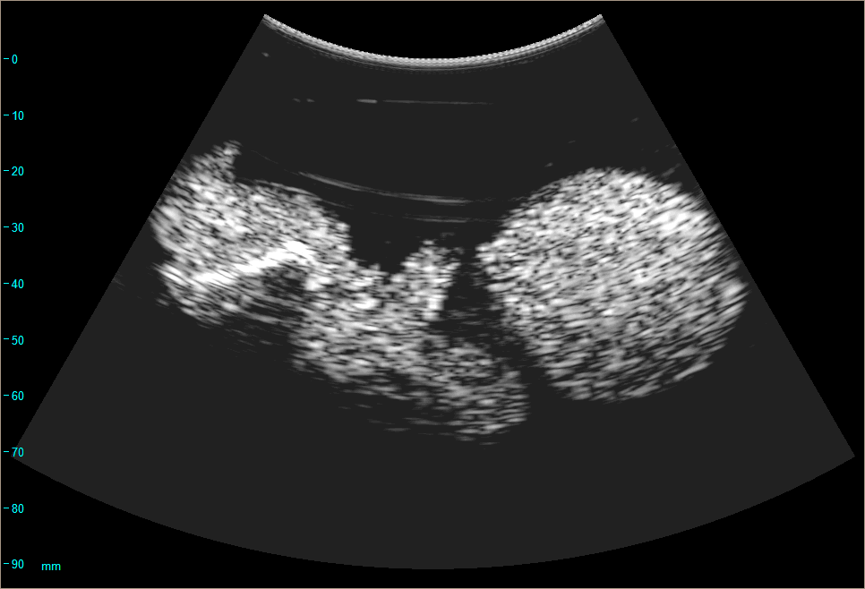
After selecting suitable measurement parameters and the optimal setting of the ultrasound image of the fetus, this is measured. Based on the determined fetometric parameters (crown rump length: approx. 105 mm, occipitofrontal andbiparietal diameter: approx. 44 mm and 30 mm, head circumference: approx. 118 mm, femur length: approx. 25 mm), the age of the fetus ist estimated to be about 17 weeks.Below the femora, dark areas may appear in the ultrasound image. These acoustic shadows are an example of artifacts in sonograms, which must be considered in their evaluation.
| Art.No. | Description |
|---|---|
| 10410 | Ultrasonic echoscope GS200i |
| or | |
| 10412 | Ultrasonic B-scan device Gi210 |
| 10430 | Ultrasound fetal phantom |
| PHY01 | Basics of pulse echo method (A-scan) |
| PHY08 | Ultrasound B-scan |
| MED07 | Ultrasound test phantom |
| MED09 | Mamma sonography |
| MED10 | Gallbladder ultrasound |