Article No. VK-MED04
MED04 Biometry at the eye phantom
Measurement of times of flight at an eye phantom as a typical biometric ultrasonic application
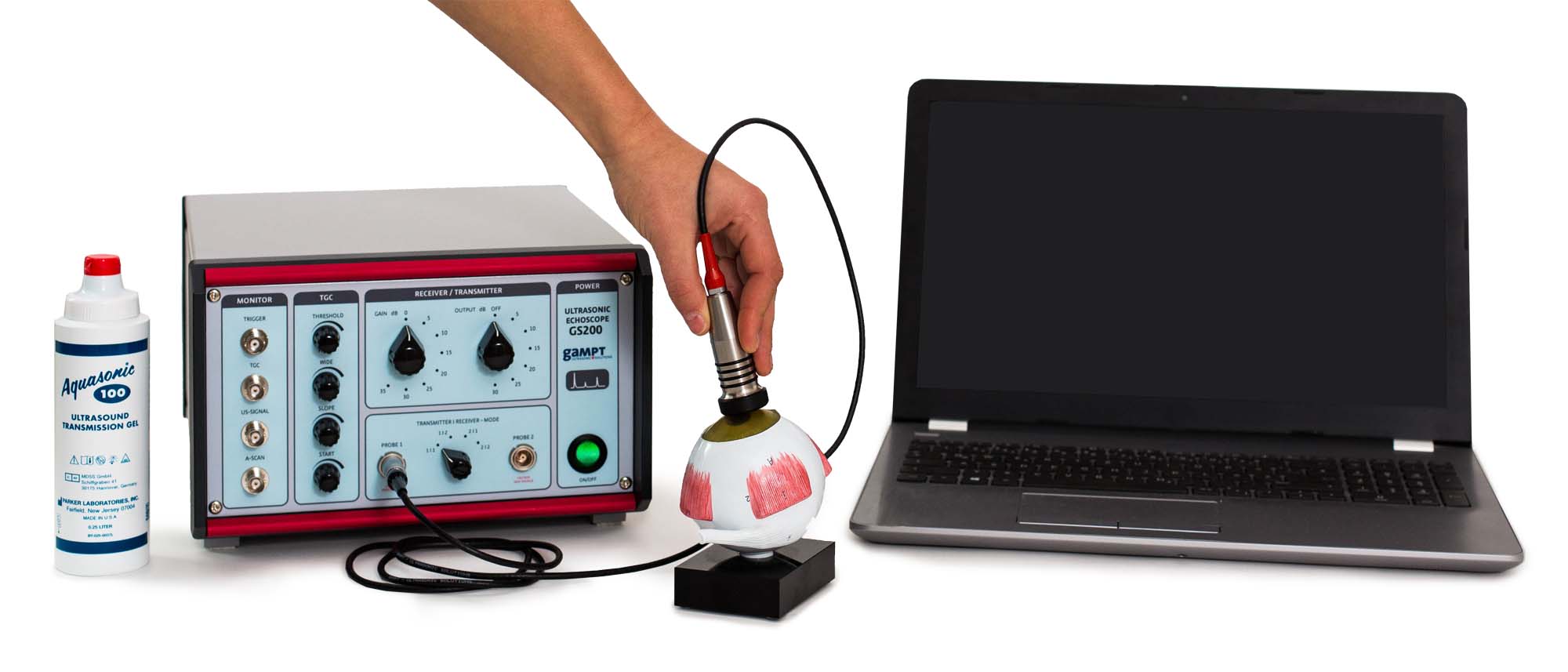
The measurement of times of flight of ultrasonic signals at an eye phantom at an enlarged scale demonstrates a typical biometric ultrasonic application based on the A-Scan method in medical diagnostics in ophthalmology.
Keywords: Ultrasonic echography, pulse echo method, time of flight, velocity of sound, reflection and transmission coefficient, A-scan, sonography of the eye, biometry
Ophthalmology is another area of medicine in which ultrasound is used. Here, ultrasound is especially important for the biometric surveying of the eye, i.e. the measurement of distances in the eye. For example, the distance between cornea and iris is very important for the calculation of the characteristics of an artificial lens, such as is implanted for patients with cataracts. Because the cornea or lens is too cloudy for optical methods, it is here necessary to use ultrasonic methods. Although new methods with laser light and the ultrasonic B-Scan method are now used, time of flight measurements of the ultrasonic echoes of an A-Scan at the eye offer a simple way to measure the eye. When calculating the sound paths from the measured times of flight it is to be noted that different sound velocities occur in the cornea, the lens, the vitreous humour and in the other areas of the eye. In the eye phantom provided, the sound velocity in the lens is around 2500 m/s and in the vitreous humour it is around 1410 m/s.
Echo signals at the eye phantom and their origins
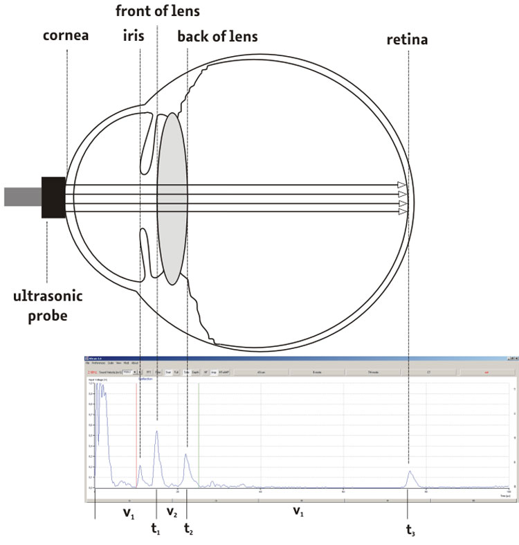
The illustration shows a schematic presentation of the eye phantom and an A-Scan image recorded with the measurement software. The individual ultrasonic echoes are here assigned to the sites of their origin in the eye phantom.
| Ord.no. | Description |
|---|---|
| 10400 | Ultrasonic echoscope GS200 |
| 10152 | Ultrasonic probe 2 MHz |
| 10222 | Eye phantom |
| 70200 | Ultrasonic gel |