Article No. VK-MED05
MED05 Vascular Ultrasound (Angiology)
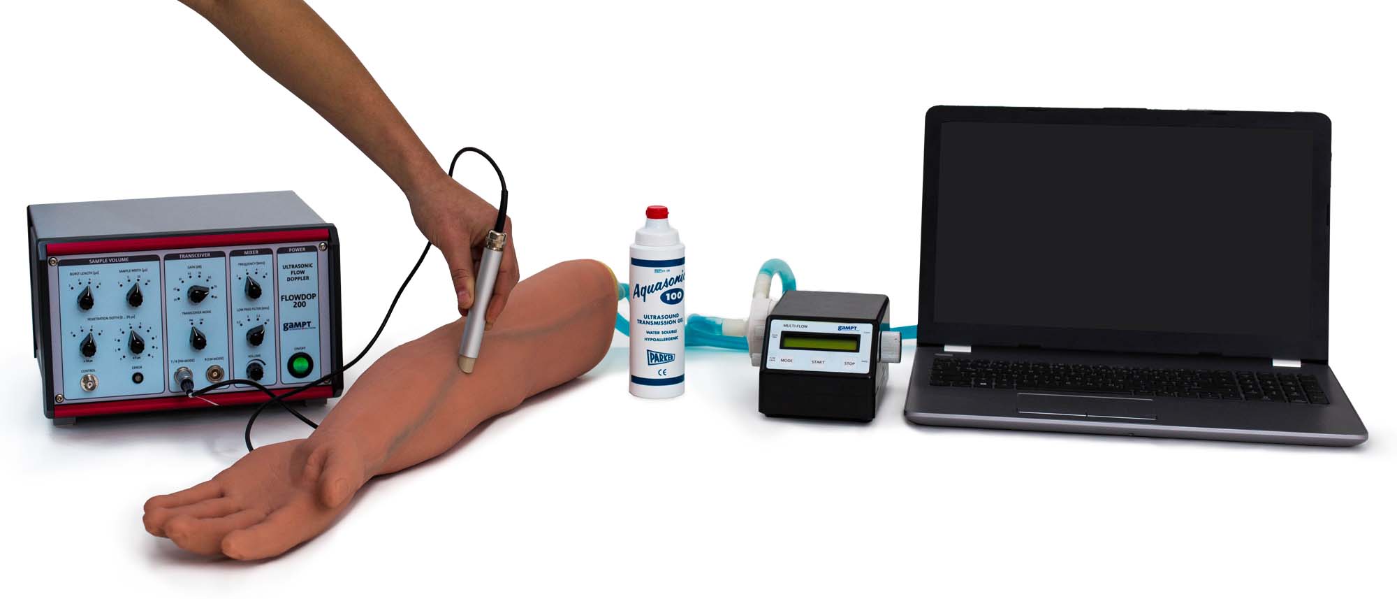
Demonstration of blood flow examinations by means of the ultrasonic Doppler method using a realistic arm phantom, Presentation of the differences between continuous venous and pulsatile arterial flow
The experiment demonstrates the performing of blood flow examinations by means of the ultrasonic Doppler method. The differences between continuous venous and pulsatile arterial flow are presented using a realistic arm phantom. Furthermore, the influence of a stenosis and the air chamber function on pulsatility is investigated.
Keywords: Ultrasonic scattering, frequency shift, Doppler effect, Doppler sonography, continuity equation, pulse curves, stenosis, air chamber function
Doppler sonography is based on the effect of the frequency shift between the sent and the received ultrasonic signal in a sender-transmitter system in which transmitter and receiver are moving in relation to each other. Using the Doppler effect, moving structures, such as e.g. flowing blood, can be investigated and their relative velocities determined and visualised.In the experiment, blood flow is investigated by means of a Doppler probe at a realistic arm phantom. A narrowing of the vessels is built into the arm phantom to simulate the influence of a stenosis. In this way, differences between healthy and altered vessels can be clearly presented in the spectral image.In addition to continuous mode the pump used can also be operated in a pulsatile mode to simulate arterial blood flow. In the experiment, the influence of the stenosis and air chamber function upon pulsatility is investigated.
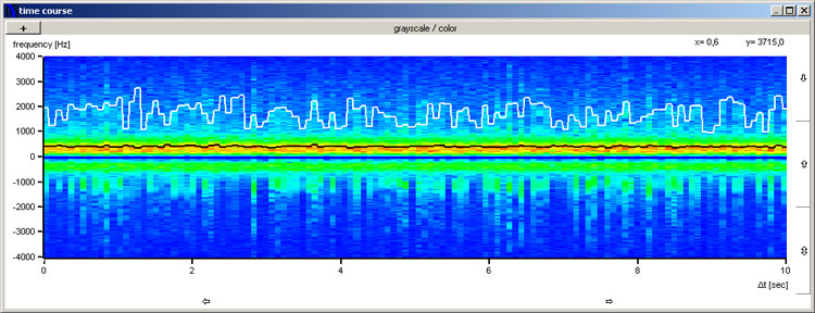
Continuous venous blood flow

Turbulent blood flow in the region of a stenosis
The measurement software provides different windows for the analysis and visualisation of the recorded Doppler signals. The example screen shots of the measurement software show in comparison the typical Doppler spectral images for a continuous venous flow (Fig. 4) and the flow in the area of a stenosis (Fig. 5).
| Ord.no. | Equipment |
|---|---|
| 50400 | Ultrasonic Doppler FlowDop200 |
| 50130 | Centrifugal pump MultiFlow |
| 50435 | Ultrasonic Doppler probe |
| 50160 | Arm phantom |
| 70200 | Ultrasonic gel |