Article No. VK-30850
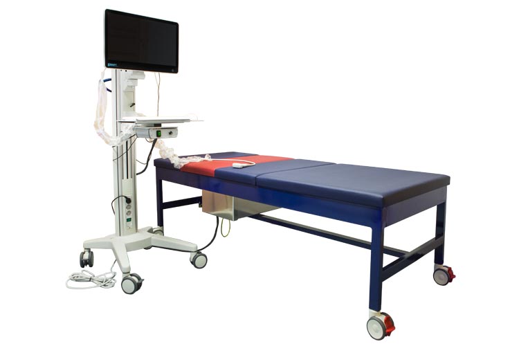
Time harmonic elastography – THED III Set
For in vivo and non-invasive palpation of the heart in real time. The resulting elasticity markers identify diastolic dysfunction. incl. delivery of an ultrasound probe of your choice (curved array, phased array)
![]()
THED research – Quantifying pathological and physiological changes through deep palpation
THED research is a pioneering research platform for the establishment of time-harmonic elastography (THE) as an innovative, MRI-validated ultrasound modality. The method enables high-precision, quantitative analysis of the shear wave velocity in deep tissue layers – up to a penetration depth of 13 cm. THED research thus opens up new diagnostic possibilities, even in challenging conditions such as ascites or obesity.
This technology was developed jointly by Charité – Universitätsmedizin Berlin and GAMPT mbH in Merseburg.
Learn more about the application of the system, especially in liver diagnostics, in our introductory video.
For scientific questions, please contact Prof. Dr. Ingolf Sack from Charité Berlin and for acquisition and support, please contact Dr. Michael Schultz from GAMPT mbH.
THED research – Precise in vivo measurement of tissue stiffness using temporal harmonic waves
Continuous Stimulation Instead of Pulsed Waves
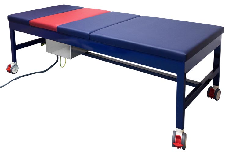
THED research was developed to reliably measure tissue stiffness in vivo in a wide range of diagnostic applications. The underlying principle differs fundamentally from conventional elastography methods: Instead of individual pulses, THED uses continuous, time-harmonic waves generated by an external vibration source. This is integrated directly into the patient bed and ensures uniform tissue stimulation over longer periods of time.
A comparison that illustrates the difference
Dr. Richard G. Barr from Northeastern Ohio Medical University described this innovative approach in the journal Radiology (May 2018, DOI link) with a vivid comparison:
“[Conventional] techniques are similar to throwing a stone into water – the waves only propagate briefly before they subside. […] Time-harmonic elastography, on the other hand, is like a bowl of water during an earthquake – the waves persist and cover a much larger area.”
Reliable Results in All Patient Groups
Dr. Barr also points out:
“[The method] enabled reliable measurements in all patient groups, including measurements down to 14 cm depth. With existing ultrasound techniques, however, high body mass index or the necessary distance to the liver capsule often lead to unreliable results.”
Sensitive and Consistent Measurements Across Organs
Thanks to continuous, frequency-controlled tissue stimulation, THED research delivers particularly sensitive and consistent measurement results for many diseases that are associated with altered biomechanical properties. The system has already been successfully used for the examination of abdominal organs such as the liver, kidney, spleen, pancreas and aorta. Initial pilot studies also show promising results in the prostate, uterus, heart and even the brain.
Seamless Comparison with MRI: A Breakthrough in Elastography
The ability of THED to generate high-resolution full-field elastograms even under difficult conditions – such as obesity or ascites – is particularly relevant for everyday clinical practice. Another decisive advantage: as stimulation takes place at precisely defined frequencies, the measured stiffness values are directly comparable with those of magnetic resonance elastography (MRE) – the current gold standard. This direct comparability allows a seamless transfer of diagnostic threshold values between ultrasound and MRI – a unique feature in modern elastography.
Enhanced Diagnostics through Combined Imaging and Deep Scanning
THED research combines high-resolution real-time imaging with precise remote scanning of organs to significantly increase the diagnostic value. With a maximum penetration depth of 13 cm, the system extends the range of conventional ultrasound technology and enables the reliable measurement of deeper organs.
Advanced Elastogram Generation Using Standard Hardware
Elastograms are generated across the entire field of view using a specially developed alias algorithm. This means that standard ultrasound hardware can be used – in compliance with all thermal and mechanical safety limits.
Versatile Viscoelastic Tissue Analysis Methods
The system offers both one- and two-dimensional methods for viscoelastic tissue analysis. Specific parameter sets are available for the examination of the heart and liver, while a universal standard parameter set supports the analysis of other organs.
Improved Spatial Orientation through Image Overlay
A semi-transparent overlay of the elastograms with the classic B-mode image facilitates spatial orientation and improves diagnostic reliability. In addition, the parallel display alongside the B-scan supports intuitive 2D guidance during the examination.
A Future-Oriented Solution for Elastography Research
THED research is aimed at research institutions working on the further development of elastographic processes and devices. With its powerful functionality and innovative design, the system offers a forward-looking solution for overcoming the current limitations of elastography.
The video shows the application of THED research to the liver. A demonstration of THED research can be arranged at our clinical partner in Berlin.
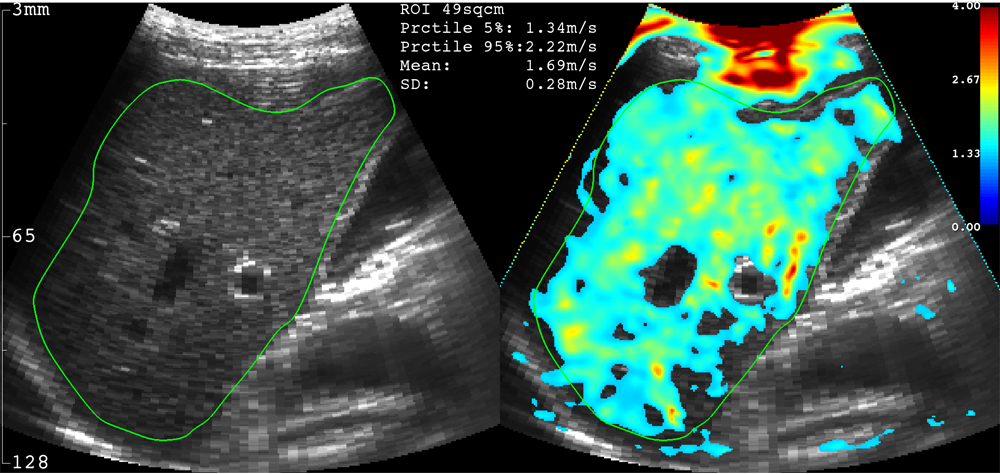
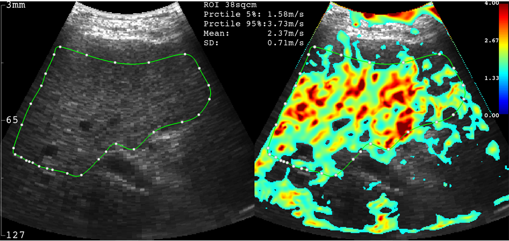
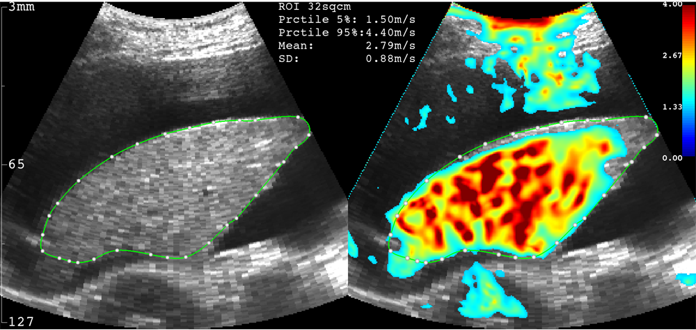
The following figures show the side-by-side display of the B-mode ultrasound image and the large B-mode elastogram on the THED computer screen.
 |
 |
 |
|
Healthy liver |
Liver with fibrosis |
Liver with fibrosis and ascites |
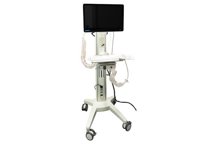
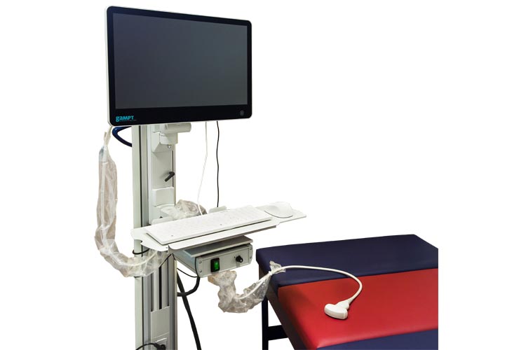
High-end ultrasound system SonixTablet (Analogic Ultrasound) in combination with software and hardware for elastography
Highly innovative ultrasound research device for elastography based on B-mode-guided motion estimation and time-harmonic waves caused by an actuator in bed form
The product THED is a joint development of Charité Berlin and GAMPT mbH in Merseburg.
If you have any questions, please contact Prof. Ingolf Sack (Charité Berlin).
![]()
Waiver of rights
The THED research system from GAMPT mbH is intended exclusively for research purposes. THED research has no intended purpose as required for medical devices in MDD 93/42/EEC and Regulation EU 2017/745. It does not comply with the general requirements of these directives. GAMPT declines any responsibility if THED research is used as a decision support system for the diagnosis of diseases or their exclusion.
News from the archive
New real-time ultrasound method developed to measure cerebral rigidity
Non-invasive real-time measurement of cerebral rigidity with THED
THED research enables the first non-invasive real-time measurement of cerebral stiffness (CS) using trans-temporal time-harmonic elastography. Cerebral stiffness describes the biomechanical environment in which neurons grow and function – a crucial factor for both brain health and neurological disease progression.
While long-term changes in CS in chronic diseases and with age have already been documented, little is known about short-term, pressure-related changes. Conventional methods such as magnetic resonance elastography or direct pressure measurements are either invasive, slow or costly.
Study on rapid, non-invasive CS measurement and initial results
A rapid, non-invasive method for measuring CS in vivo has now been tested in a recent study. Continuous external shear wave stimulation in the frequency range of 27-56 Hz was used. The generated shear waves were detected by trans-temporal ultrasound and converted to shear wave velocity (SWS) maps using multi-frequency wave inversion – an established parameter for estimating tissue stiffness.
The results show:
- an average cerebral SWS of 1.56 ± 0.08 m/s,
- a significant age-dependent decrease in CS (R = -0.76, p < 0.0001),
- a direct influence of the Valsalva maneuver on CS with an increase in SWS of 10.8% (p < 0.0001) – in response to increased intracranial pressure.
These findings indicate that cerebral stiffness is suitable as a potential surrogate marker for intracranial pressure – with the major advantage that it can be recorded non-invasively, in real time and cost-effectively in the future.
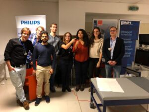
THED at the EUROSON Training Course on Liver Elastography in Timisoara
In November 2018, THED research participated in the EUROSON Training Course on Liver Elastography, organized by the Ultrasound Learning Centre in Timisoara.
Around 40 physicians and medical researchers took the opportunity to test and compare various ultrasound systems and elastographic techniques — including our research system, THED — in a hands-on setting.
The course also marked the launch of a pilot study on 2D time-harmonic elastography, initiated at the Victor Babeș University of Medicine and Pharmacy in Timisoara. We are proud to be part of this promising initiative that brings new momentum to liver diagnostics and research.
 |
 |
New Imaging Technique for Liver Diagnostics in Overweight Adolescents
Researchers at Charité – Universitätsmedizin Berlin have successfully tested an innovative method for examining liver disease in overweight adolescents.
The technique, known as Time-Harmonic Elastography (THE), allows for an accurate assessment of disease severity without the need for invasive liver biopsies. By non-invasively measuring liver stiffness, THE provides a reliable evaluation of disease progression — a significant advantage, especially for young patients.
The promising results have now been published in the renowned journal Radiology.
Links:
https://idw-online.de/de/news697547
https://www.eurekalert.org/pub_releases/2018-06/c-ub-nrp061818.php
Ultrasound Time-Harmonic Elastography: Detecting Liver Fibrosis in Adolescents with Severe Obesity and NAFLD
Pediatrician Dr. Christian Hudert and colleagues at Charité – Universitätsmedizin Berlin have published a study highlighting the diagnostic potential of time-harmonic elastography (THE) for identifying liver fibrosis stages in overweight to severely obese pediatric patients with non-alcoholic fatty liver disease (NAFLD).
The study demonstrated that THE was feasible in 100% of cases, with no technical failures, even in patients with extreme obesity. These findings suggest that THE could serve as a reliable, non-invasive alternative to liver biopsy in the future — offering a safer and more patient-friendly approach to fibrosis assessment in children and adolescents.
THED enables non-invasive assessment of the kidney
THED research is a cutting-edge research platform designed to establish MRI-validated Time-Harmonic Elastography (THE) as an advanced ultrasound imaging modality.
The system provides quantitative analysis of shear wave speed at depths of up to 13 cm, enabling reliable wave propagation even beyond fluid accumulations (e.g., ascites) and allowing for the accurate assessment of obese patients.
A new publication by the research group at Charité – Universitätsmedizin Berlin now demonstrates the successful application of THED in the kidney, further expanding the system’s potential in clinical diagnostics.
Full-field-of-view time harmonic elastography of the native kidney
Marticorena Garcia SR, Großmann M, Lang ST, Nguyen Trong M, Schultz M, Guo J, Hamm B, Braun J, Sack I, Tzschätzsch H. Ultrasound in Medicine and Biology 2018